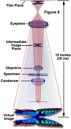
Direct light and reflected or diffracted light are brought together in phase peaks valleys match to form an image of the specimen. Objective lenses in a light microscope.

Molecular Expressions Microscopy Primer Anatomy Of The Microscope Magnification
Project the magnified image of the specimen onto the plane of the.
. Most light microscopes are compound microscope that contains at least two lenses. The light rays which reflect off of the object are then focused into a magnified image. The power of the light microscope is limited by the wavelength of light and can magnify something up to 2000 times.
These microscopes generate images at very. The picture appears on a monitor. A light microscope can magnify images up to 1000x while electron microscopes can even magnify images more than 100000x Nester 2001.
16 Atomic force microscope The AFM is one of the foremost tools for imaging measuring and manipulating matter at the nanoscale. Some modern instruments that dont contain lenses are still known as microscopes because they magnify objects. Electron microscopes on the other hand can produce much more highly magnified images because the beam of electrons has a smaller wavelength which creates images of higher resolution.
It does this by creating a magnified image through the use of a series of glass lenses which first focus a beam of light onto or through an object and convex objective lenses to enlarge the image. Types of light microscopes. It does this by creating a magnified image through the use of a series of glass lenses which first focus a beam of light onto or through an object and convex objective lenses to enlarge the image.
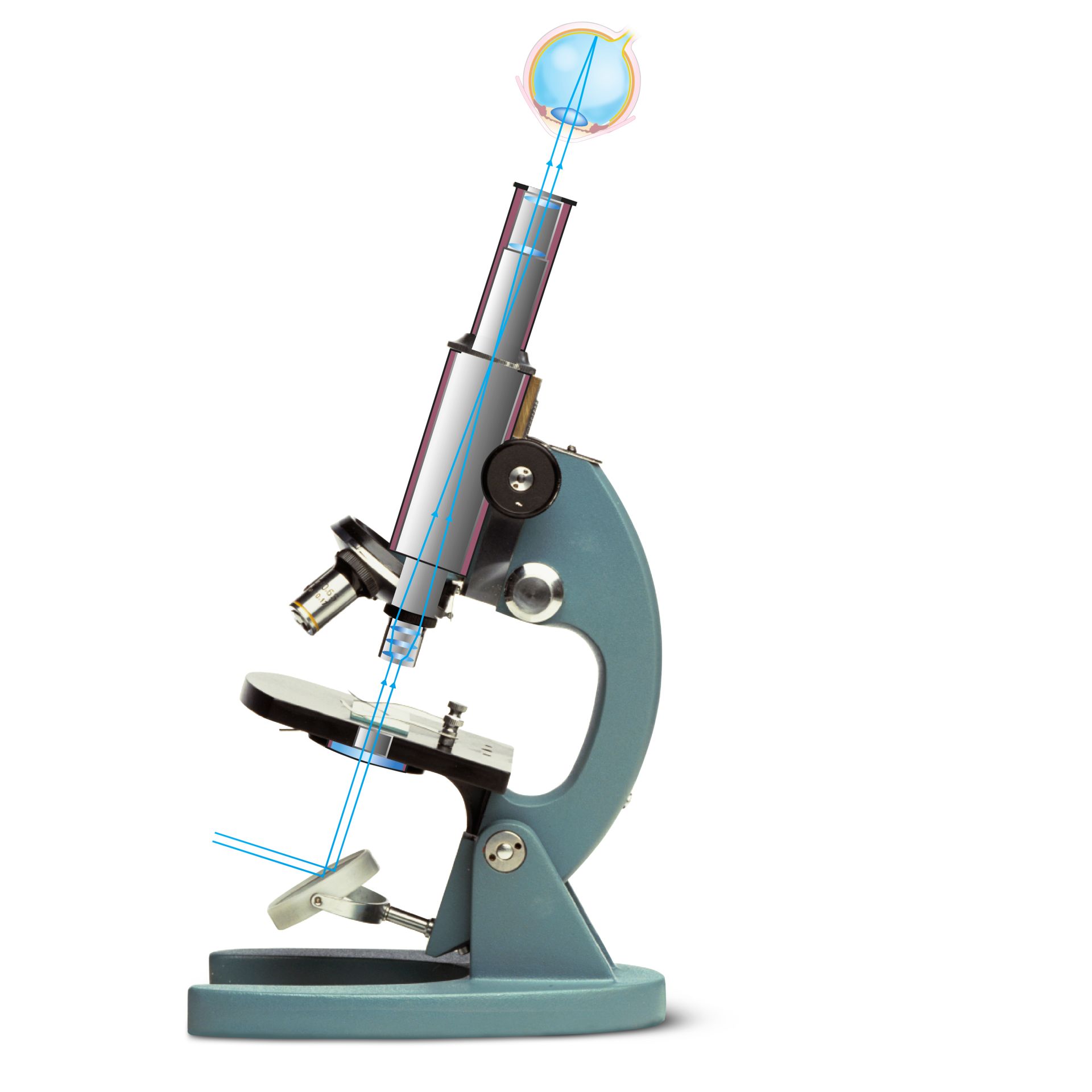
This light is refracted and focused by the lens to produce a virtual image on the retina. These pages will describe types of optics that are used to obtain contrast suggestions for finding specimens and focusing on them and advice on using measurement devices with a light microscope. This microscope is used to exam internal structures of living microorganisms.
Focus the image of specimen on the objective focal plane D. Describe How A Light Microscope Creates A Magnified Image. Describe how a light microscope creates a magnified image.
Its an upright microscope that produces a two-dimensional image and has a higher magnification than a stereoscopic microscope. Prepare wet mounts of cheek cells. They use lenses to focus light on the specimen magnifying it thus producing an image.
Do not create a magnified image of the specimen E. In the case of a SEM you do not see light but. Calculate the total magnification and field of view for the lenses of an optical microscope.
The light microscope is an instrument for visualizing fine detail of an object. The microscope must accomplish three tasks. Magnify the image that has been focused on the ocular focal plane B.
Focus the light from a bright source onto the specimen C. A simple microscope or magnifying glass lens produces an image of the object upon which the microscope or magnifying glass is focused. This type of microscopy is a Phase-Contrast using two light beams passing through prisms.
For example the scanning tunnelling microscope and the atomic force microscope measure the shape of a surface by measuring the distance between the microscopes probe and the surface. A light microscope is a biology laboratory instrument or tool that uses visible light to detect and magnify very small objects and enlarge them. A light microscope is an instrument that creates a clear magnified image through a series of lenses.
The bright field microscope is best known to students and is most likely to be found in a classroom. Learning Objectives Upon completion of this laboratory you will be able to. Outline the components of an optical microscope.
What you see when you look through a light microscope is a magnified image made from light reflecting off an object. The specimen is normally placed close to the microscopic lens. As the beam scans the surface of the sample a highly magnified image is created which allows the system operator to view the samples microscopic features clearly.
Light Microscopy HISTOLOGY AND CYTOLOGY MODULE Histology and Cytology Notes 2 LIGHT MICROSCOPY 21 INTRODUCTION Microscopes are instruments designed to produce magnified visual or photographic images of objects too small to be seen with the naked eye. The microscope is an optical instrument that uses a lens or a combination of lenses to produce magnified image of small object. Describe what the differences are.
The light microscope is an instrument for visualizing fine detail of an object. Examine prepared slides under scanning low high and oil immersion lenses. Describe field of view and depth of field.
A light microscope focuses a light source at a specimen through a series of lenses. A compound light microscope is a type of light microscope that uses a compound lens system meaning it operates through two sets of lenses to magnify the image of a specimen. Webb in Encyclopedia of Food Sciences and Nutrition Second Edition 2003 Principles.
Produce a magnified image of the. The microscope is able to create a 3-D picture of the specimen based on the way the electrons bounce off it. The light ray from a mirror is reflected through the object or specimen into the objective lens which produces first magnification.
Anatomy Of The Microscope The Concept Of Magnification Olympus Ls

Light Microscopes An Overview Sciencedirect Topics
How Does An Optical Microscope Work Dk Find Out


0 Comments